2014_01 Glimpse into the Nanoworld
What is the Topic?
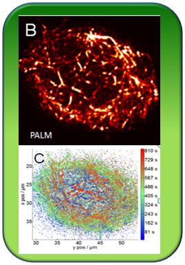
Electron microscopy provided a great breakthrough half a century ago, because the invisible details of cells became visible for the first time. Electron microscopy requires to kill the specimen, cut it into ultrathin sections and to observe it under vacuum. For life-cell observations until recently the magic Limit was 0.3 µm (1 micron, µm, corresponds to 1/1000 mm). Using a new microscopical technology, called PALM, allows to break this limit and to penetrate into the nanoworld. Together with the team of Prof. Dr. Uli Nienhaus in the Institute of Physics of the KIT our team members Dr. Steffen Durst and Dr. Jan Maisch succeeded to administer PALM-Microskopy for the first time to plant cells and this even in the third dimension. This allowed to image with 20 nm precision (1 Nanometer, nm, corresponds to a millionth of a mm) a basket-like net consisting of the muscle protein actin that encages the plant nucleus and guides the nucleus to the correct site in the cell.
Publication
103. Durst S, Hedde PN, Brochhausen L, Nick P, Nienhaus GU, Maisch J (2014) Organization of perinuclear actin in live tobacco cells observed by PALM with optical sectioning. J. Plant Physiol 141, 97-108 - pdf

