
Microtubules in action
Publication
Himmelspach, R., Wymer, C.L., Lloyd, C.W., Nick, P. (1999) Gravity-induced reorientation of cortical microtubules observed in vivo. Plant J. 18, 449-453 (54 quotations, state 27.12.2015) - pdf
What is the topic?
Subtending the plasma membrane of plant cells parallel bundles of microtubules are observed. These so called cortical microtubules serve as tracks for the movement of small rosettes consisting of cellulose synthase, the enzyme that produces cellulose fibers. The cellulose synthase is pulled by kinesin motors and leave at the outer face of the membrane a trace, the microfibrils, fibers made from cellulose. The direction of these fibers determines the axis of growth for plant cells - expansion will always occur perpendicular to the fiber direction. Cortical microtubules can change their orientation in response to environmental signals such as light, gravity or touch and this will change then the axis of cell growth. This allows plants to adjust their shape to the environment. This phenomenon was discovered during investigations on phototropism, the bending of seedlings towards light (Nick et al. 1990 - more...).
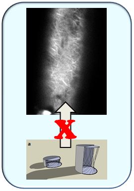
But how can microtubules change their orientation? Are they actually turned or do they disassemble and reassemble in a new direction? To visualise microtubules, so far fluorescently labelled antibodies against tubulin were employed, which required chemical fixation (which will kill the cell of interest). Thus, only still images of the final situation of the reorientation process could be collected. To get from still images towards a movie, in this work tubulin was purified from pig brains, labelled fluorescently and then microinjected into living cells of intact maize seedlings. This was technically very challenging, since one has to hit an extremely small rim of cytoplasm just underneath the cell membrane. The labelled tubulin was integrated into the microtubules of the hosting maize cell and thus rendered these microtubules visible. Now, we could stimulate the cell by gravity and watch the reorientation of microtubules at the upper surface. This allowed, for the first time, to see the transition between the non-stimulated initial phase and the completed response of microtubules. The transition consisted in microtubules that were oriented criss-cross, which was clear evidence against the hitherto prevailing model that assumed that microtubules turned. Instead, they disassemble individually and are then reassembled in the new orientation.