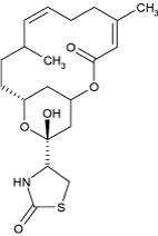
Latrunculin B
Source
red-sea sponge Latrunculia magnifica
Mode of action
Latrunculin B sequesters G-actin at a 1:1 ratio (Coué et al. 1987). It therefore eliminates actin filaments depending on their innate turnover. It is not “disrupting” actin filaments – a stable filament without turnover will not be affected. Binding at the barbed end causing a block of both association and dissociation. Dissociation at the pointed end continues. Most potent actin inhibitor, because activity is irreversible.
Binding site
The binding site of Latrunculin A has been experimentally determined by steric hindrance of thymosin and modelling (Morton et al. 2000) to be at the interface of monomers. The exact binding pocket for the similar Latrunculin B has been derived from structural modelling (Banting and Clark 2012). Details see in the data sheet.
Practical aspects
Kd of binding is 0.2 µM. Solubility in water is low, stock solutions in DMSO (25 mg/mL) and ethanol (25 mg/mL) can be used, store at -20°C.
- Arabidopsis seedlings: 1 µM cause a complete block of cell growth (Baluška et al. 2001)
- Rice coleoptiles: 1-10 µM of Lat B for 1 hour block gravitropic curvature and lateral auxin transport (Nick et al. 2009).
- BY-2: 2 µM cause rapid intracellular clustering of pin1 (Chang et al. 2011). 65 nM alter division synchrony (Durst et al. 2013). 100 nM block proliferation (Durst et al. 2014). 500 nM overnight eliminate actin (Maisch et al. 2009), block endocytosis and uptake of Trojan Peptoids and CPPs (Eggenberger et al. 2009, 2011). 10 µM for 1-2 h block nuclear migration (Frey et al. 2009; Klotz and Nick 2012). Protoplasts: 1 µM modulates regeneration (Zaban et al. 2013) 1 µM to see immediate response under the microscope (Gao et al. 2016)
- Vitis: 2 µM 30 min modulate elicitor triggered alkalinisation (Chang and Nick 2012), 1 µM 30 min alter defence genes (Qiao et al. 2010). protoplasts: 1 µM for 30 min changes volume control (Liu et al. 2013) 2 µM to see effect on cell death (Chang et al. 2015)
- Plasmopara: 10 nM affect germ tube formation (Riemann et al. 2002)
Literature
- Mode of action: Coué M, Brenner SL, Spector I, Korn ED (1987) Inhibition of actin polymerization by latrunculin A. Fed Europ Biochem 213, 316-318 - pdf
- Effect on plants: Baluška F, Jasik J, Edelmann HG, Salajová T, Volkmann D (2001) Latrunculin B-Induced Plant Dwarfism: Plant Cell Elongation Is F-Actin-Dependent. Developmental Biology 231, 113-124 - pdf
- Binding site (Latrunculin A): Morton WM, Ayscough KR, McLaughlin PJ (2000) Latrunculin alters the actin-monomer subunit interface to prevent polymerization. Nature Cell Biol 2, 376-378 - pdf
Publications from our group using Latrunculin B
35. Riemann M, Büche C, Kassemeyer HH, Nick P (2002) Microtubules and actin microfilaments guide the establishment of cell polarity during early development of the wine pathogen Plasmopara viticola. Protoplasma 219, 13-22
67. Maisch J, Fišerová J, Fischer L, Nick P (2009) Actin-related protein 3 labels actin-nucleating sites in tobacco BY-2 cells. J Exp Bot 60, 603-614
71. Frey N, Klotz J, Nick P (2009) Dynamic bridges - a calponin-domain kinesin from rice links actin filaments and microtubules in both cycling and non-cycling cells. Plant Cell Physiol 50, 1493-1506
73. Eggenberger K, Schröder T, Birtalan E, Bräse S, Nick P (2009) Passage of Trojan Peptoides into Plant Cells. ChemBioChem 10, 2504-2512
75. Nick P, Han M, An G (2009) Auxin stimulates its own transport by actin reorganization. Plant Physiology 151, 155-167
81. Qiao F, Chang X, Nick P (2010) The cytoskeleton enhances gene expression in the response to the Harpin elicitor in grapevine. J Exp Bot.61, 4021-4031
82. Eggenberger K, Mink C, Wadhwani P, Ulrich AS, Nick P (2011) Using the peptide BP100 as a cell penetrating tool for chemical engineering of actin filaments within living plant cells. ChemBioChem 12, 132-137
87. Chang X, Heene E, Qiao F, Nick P (2011) The phytoalexin resveratrol regulates the initiation of hypersensitive cell death in Vitis. PLoS ONE 6, e26405.
88. Klotz J, Nick P (2012) A novel actin-microtubule cross-linking kinesin, NtKCH, functions in cell expansion and division. New Phytologist 193, 576-589
91. Chang X, Nick P (2012) Defence Signalling Triggered by Flg22 and Harpin Is Integrated into a Different Stilbene Output in Vitis Cells. PLoS ONE 7: e40446
93. Zaban B, Maisch J, Nick P (2013) Dynamic actin controls polarity induction de novo in protoplasts, J Int Plant Biol 55, 142–159
97. Durst S, Nick P, Maisch J (2013) Actin-Depolymerizing Factor 2 is Involved in Auxin Dependent Patterning, J Plant Physiol 170, 1057-1066
98. Liu Q, Qiao F, Ismail A, Chang X, Nick P (2013) The Plant Cytoskeleton Controls Regulatory Volume Increase. BBA Membranes 1828, 2111–2120
103. Durst S, Hedde PN, Brochhausen L, Nick P, Nienhaus GU, Maisch J (2014) Organization of perinuclear actin in live tobacco cells observed by PALM with optical sectioning. J Plant Physiol 141, 97-108
114. Chang X, Riemann M, Nick P (2015) Actin as deathly switch? How auxin can suppress cell-death related defence. PloS ONE, doi: 10.1371/journal.pone.0125498
119. Gao N, Wadhwani P, Mühlhäuser P, Liu Q, Riemann M, Ulrich A, Nick P (2016) An antifungal protein from Ginkgo biloba binds actin and can trigger cell death. Protoplasma, DOI 10.1007/s00709-015-0876-4

